Once again courtesy of Not Exactly Rocket Science comes an article about a rather bizarre (to us at least) factor in the process of arthropod molting: apparently, they also shed the lining of their lungs while they’re at it.
Now, this is a little bit different from what we might imagine (yeah, like discarding your entire skin at once to emerge bigger is nothing odd.) Insects – and arachnids, and crustaceans, and so on, the whole class of Arthropoda – don’t have lungs anything like the mammals; instead, the vast majority of them have little holes along the sides of their thoraxes and abdomens, called spiracles, that feed air more directly into the tissues through tracheae (or tracheoles – I’m not sure which is proper,) as partially illustrated before. When molting, the entire lining of these passages is pulled out and discarded as well, interrupting their breathing for several minutes – according to the article, they increase their respiratory rate ahead of time, flooding their system with oxygen, then breathing halts during the molt, following which they start gulping air again. Not surprising, really. The respiratory rate on arthropods is typically pretty low anyway, which is why spiders can survive under water for extended periods solely on the air that clings to their bodies.
The image they used to illustrate this for the article wasn’t terribly enlightening, and I wondered if I had something more useful in my stock. I’d seen the little white threads left behind within the molted chitins, but always assumed they were tendons or something. I was in the process of checking out my arthropod stock images when I suddenly remembered: I did have access to a recently molted exoskeleton – one of the better types to use for such illustrative purposes, in fact.
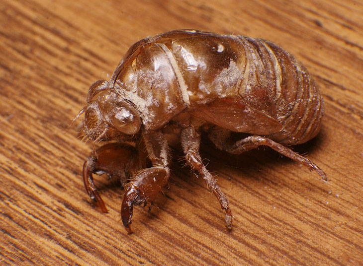
Cicadas are a common insect at this time of year throughout the US, and as they emerge from a subterranean existence and molt into reproducing, flying adults, the wonderfully menacing brown skins left behind on treetrunks and fenceposts are often not hard to find. As molted exoskeletons go, they’re large, sturdy, and hardened into shape (one prolific year as I was growing up in central NY, I collected over a dozen in one session and perched them all on a styrofoam cooler on our porch, convincing my mother momentarily that we were being infested.) I had just spotted one the other day on our fence, so I trotted back outside and brought it in for a studio session.

I will pause here for moment to reflect on body shapes. The adult cicada doesn’t look very far removed from this, though the head is broader, but it’s easy to mistake it for something entirely different because of the wings, which stretch over twice the length of the body. The majestic clear membranes emerge entirely from those embarrassing little flaps seen here over the hindmost leg, unfolding and fleshing out in a matter of hours, making clown cars look feeble in comparison.
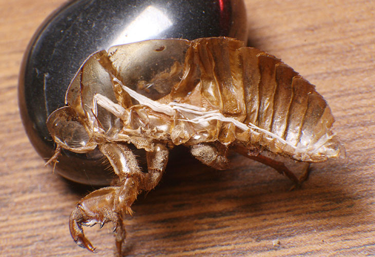
Despite the rigidity of the exoskeleton, it still took a bit of care to bifurcate it with a scalpel to reveal the interior. But once I had my cicada on the half-shell, the tracheoles were obvious white threads scattered within (the hematite stone in the background is just a bonus, what I had handy on my desk to prop up my subject.)

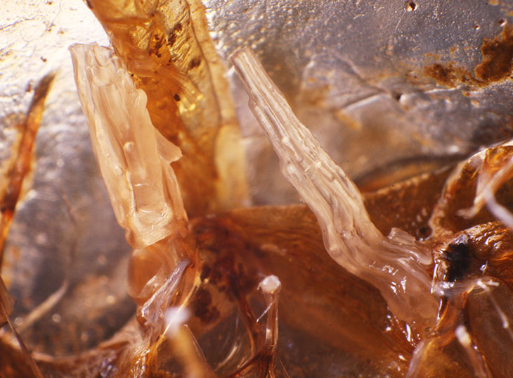
Curious to see if I could produce a better look, I soaked my subject in alcohol for a short while, knowing this is a good way to soften up chitin for a little flexibility. This kind of worked, but not as well as I’d hoped; while I could stand up the tracheoles for a little better detail, the alcohol turned them faintly translucent so the detail wasn’t as distinct as it could be.

I had no luck in trying to manipulate them to be sure, but what I think you’re seeing here is several branches clustered together; within the body, they would be split apart at the base and spreading out throughout the tissues for better oxygen distribution. These were a few millimeters in overall length, so it was hard enough to visually separate them, much less do so with the tip of a scalpel, possibly made worse by the alcohol causing them to cling together.
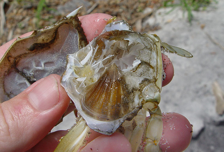
This made me remember some images, and questions, from a decade ago in Florida, when my brother and I had discovered the newly-molted chitin of a blue crab (Callinectes sapidus) – we knew it was recent because we also found the former occupant, still soft and very shy. Crustaceans, as odd as it might seem, are still arthropods, more closely related to terrestrial insects than to any aquatic neighbor. So here’s a look at the discarded exoskeleton, split open to show the interior – you’re seeing it from the left side, head towards the left of the frame.

The triangular brown mass extending from the bottom edge is one of the ‘lungs’ – the other is just barely visible edge-on at the top. I was familiar with this anatomy from having eaten crabs, but it seemed extremely peculiar that the lungs would be left behind with the molted skin; however, we had the former occupant right nearby so we were pretty sure this was not just a dead specimen. Now I know that they really do discard the lungs with the rest.
As to why this should occur, I can only begin to speculate. When developing as a fetus, nearly all species develop from a cluster of cells into a donut shape; the hole in the middle is the alimentary canal, what will become a digestive system. This does indeed make us, damn near everything really, a glorified tube. While the development of limbs from little nubs that sprout from this tube is well known, the development of the breathing apparatus takes on many forms, from the gill arches of fish to the elaborate lung systems of mammals. Somewhere in there, arthropods seem to have gotten their respiratory system closely enough linked with their exoskeleton that both are shed and developed anew with each stage of development. Weirder things happen, like the metamorphosis of many insects from pupa through chrysalis to winged adult, pretty much becoming little more than liquid in the process, but still…


